Abstract
In this study, we present a novel, high content technique using an innovative cylindrical acoustic transducer, stroboscopic fast imaging and homodyne detection to recover the mechanical properties (dynamic shear modulus) of living adherent cells at low ultrasonic frequencies. By analyzing the micro-oscillations of cells we were able to simultaneously mechanotype whole populations of cells with sub-cellular resolution. The technique can be combined with standard fluorescence imaging allowing to further cross-correlate biological and mechanical information. We demonstrate the potential of the technique by mechanotyping co-cultures of different cell types with significantly different mechanical properties.
Advanced Science, 2024
https://doi.org/10.1002/advs.202307929
Graphical Abstract:
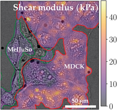
A novel, high content technique using a cylindrical acoustic transducer, stroboscopic fast imaging, and homodyne detection to recover the mechanical properties (dynamic shear modulus) of living adherent cells at low ultrasonic frequencies. By analyzing the micro-oscillations of cells this allows to simultaneously mechanotype whole populations of cells with sub-cellular resolution.
Description of setup:
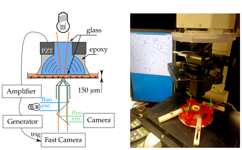
Left: Acoustic tranducer consisting of piezoelectric element (PZT) with epoxy lens focusing acoustic waves through (blue) water enclosed between upper and lower glass window. The transducer is lowered into the growth media (orange) in a petri dish until the distance between the transducer glass and petri dish is about 150 µm. Cells adhering to the petri dish are deformed by shear flow in the growth media caused by the acoustic waves. The petri dish is placed on an inverted microscope with both transmitted light illumination for fast imaging of the cells while they move in the shear flow, and reflected light illumination for fluorescence imaging. A function generator generates wave pulses that are amplified and sent to the PZT and triggers the fast camera for the stroboscopic imaging.
Right: Acoustic transducer placed in its adjustable holder (red) on the inverted microscope with the images of the cells in the petri dish displayed on the monitor behind.