
While some scientists look to the stars with telescopes in search of the origin of the universe, others turn to microscopes to understand the origin of life.
Through his PhD work at the Centre for Molecular Medicine Norway (NCMM), Lin Xue used optical microscopy to visualize how the first primitive cells could have evolved around 4 billion years ago.
What he saw not only gives us a clue as to how these primitive cells can form, but also how they may have communicated.
The first steps to creating living cells
Cells are considered the smallest unit of life. Even though they are small, the cells that make up our bodies are complex. They contain proteins, genetic material, structures known as organelles, nutrients and more.
All these components are surrounded by a cell membrane, which separates and protects the interior of the cell from the surroundings.
This complexity is the result of billions of years of evolution. The first cell-like structures to form eons ago were likely much more primitive. A fundamental step, however, would have been to create a compartment separated from the environment.
Cell membranes are mainly made up of lipids, which are molecules such as fats, that are insoluble in water. This means that when mixed with water, lipid molecules tend to stick together. It’s possible that certain conditions on the early Earth allowed lipids to assemble into a membrane, creating the first cell-like structures known as protocells.
What if we could witness these first steps of cell creation with our own eyes?
Mimicking early Earth conditions to build protocells
The goal of Xue’s PhD was to study how these protocells form and behave, to better understand how early life may have evolved. To do so, he mimicked protocell formation in the lab.
“To be able to study protocells, we make them in the lab by using simple molecules, surfaces and varying temperatures that resemble the conditions of the early Earth. We can then observe the protocells using different microscopes and study their behavior. This can give us clues as to how protocells gradually developed more complexity, and ultimately evolved into the first living cells, explains Xue.
To create protocells, Xue and colleagues would deposit a mix of simple lipid molecules in an aqueous solution on solid surfaces that reflect early Earth minerals and rocks.
Through the microscope, they could see how the lipids would come together, creating a membrane that gradually changed shape to form several closed, spherical protocells.
Protocells are connected through intricate network
When observing protocell formation, Xue and his colleagues noticed something intriguing.
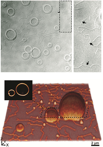
“One of our earliest observations when creating protocells, was that the lipids would not just come together to form many small protocell structures, but also a web of thin, lipid tube-like networks between the individual protocells”, says Xue.
These tubular networks between individual protocells resembled structures called tunneling nanotubes that are known to connect many different cell types that exist today. Tunneling nanotubes are used by cells to communicate with their neighbors by allowing them to transport molecules and organelles between each other.
“Seeing these tubes forming between protocells made us speculate whether they could represent one of the earliest forms of communication between cell-like structures”, explains Xue.
Another essential part of life is the ability to adapt to the environment. Once shielded from their environment by a lipid membrane, cells have evolved several ways to communicate with their neighbors and surroundings across their membranes.
By communicating with their neighbors through a tubular network, protocells could have formed colonies that were more adaptable to their surroundings. It could have allowed protocells to share for instance genetic material, and together develop the complexity needed for life.
Communication would light up the protocells
To study if the protocells could share material through the tubular network, Xue added genetic material, DNA, to the mix. The DNA was labeled with fluorescent dye that can emit light. This allowed Xue to see it with a specialized microscope.
“The DNA would end up inside the protocells. We could then see the protocells light up as shining spheres in the microscope because of the fluorescent dye attached to the DNA”, explains Xue.
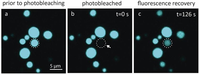
If you shine an intense light on a fluorescent dye for some time, the dye will become bleached, a phenomenon called photobleaching. This means that it no longer emits light in the microscope, and instead becomes completely dark. If you bleach a single protocell containing fluorescent DNA, it will become invisible in the microscope.
However, if the unbleached neighboring protocells are connected through a tubular network, they can pass on their fluorescent material, and the bleached protocell will eventually light up again.
“We observed that when we bleached a protocell, it would gradually light up again. This told us that the protocells were connected and able to share molecules. We even showed that they were able to pass on genetic materials between each other”, says Xue.
The creation of protocells took time and patience
The earliest records of life on Earth found to date resemble colonies of microorganisms. This raises the possibility that life evolved from colonies of protocells that teamed up, like that observed by Xue and his colleagues. Then communication through nanotubes could have been an important step towards developing the first living cells.
Exactly what happened around 4 billion years ago when the first lifeform is thought to have emerged, we may never know for sure. Nonetheless, by observing protocell formation and behavior in the microscope, Xue has gotten a glimpse of what might have happened.
“What I enjoyed the most during my PhD was sitting by the microscope for hours observing the protocells” says Xue.
“Experiments don’t always work the first time you try it, and it was a lot of trial and error to be able to create the protocells in the first place. But when it worked, and we could see these beautiful structures in the microscope, the many hours it took was all worth it.”
In a way, the origin of life was likely a result of nature's trial and error over time, which under the right circumstances and conditions eventually worked out just right.